Radiology Cases
Interesting Radiology Cases from Daily Practice and a Personal Reference
Tuesday, January 27, 2015
Abdominal Parietal Hernias at the Costal Margin
http://www.ncbi.nlm.nih.gov/pmc/articles/PMC1408328/
Tuesday, January 20, 2015
Long Plantar Ligament Sprain
? Accessory Medial Meniscofemoral Ligament
Saturday, February 9, 2013
Voice Dictation
My experience with speech recognition, including a speech (non)recognition dictionary
by Michael Tobin, MD, PhD
Article from 2002
Sit up and beg: How to train your speech recognition system
Article from 2013
Tuesday, November 13, 2012
Prefemoral Fat Pad Contusion from Direct Injury
Prefemoral Fat Pad = Posterior Suprapatella Fat Pad = Supratrochlea Fat Pad
Also note -- femoral cortcal disruption and small subperiosteal haematoma.
Saturday, September 15, 2012
Left Pudendal Neuralgia in Cyclist
Alcock's Canal
Friday, May 25, 2012
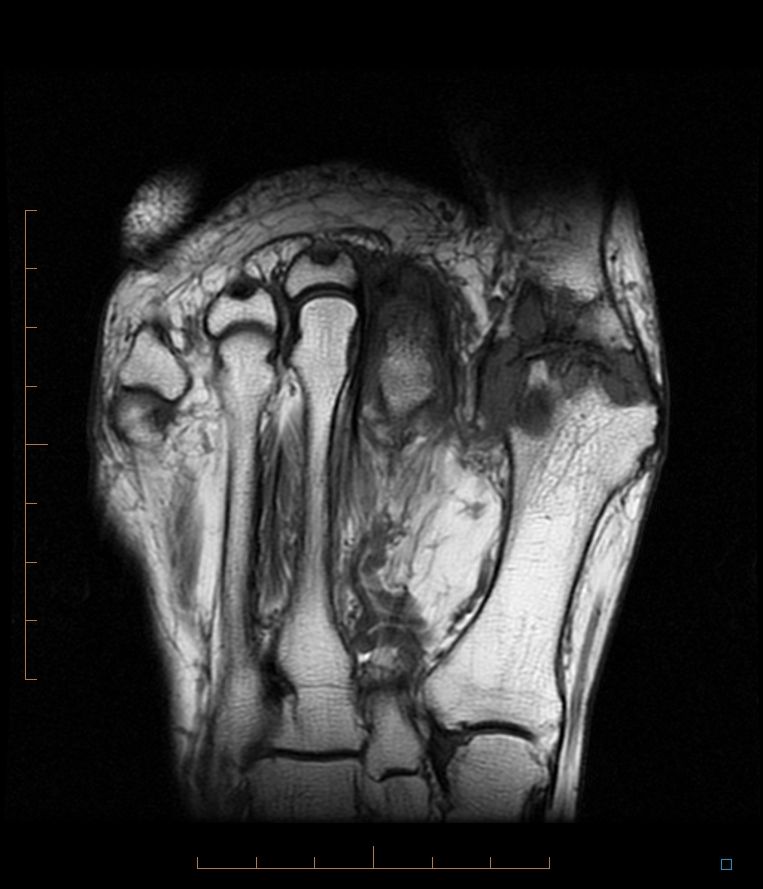
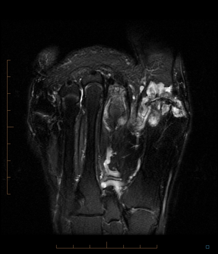
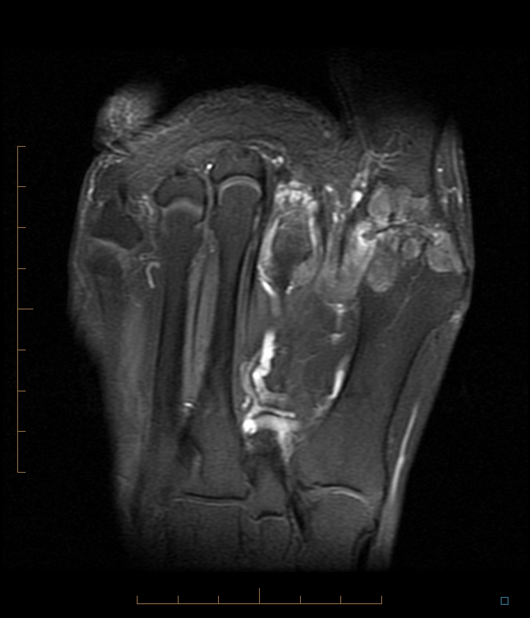
Unicameral Bone Cyst
Gout
Sunday, April 8, 2012
Baastrup Disease Thoracic Spine
Tuesday, February 28, 2012
Posterior capsular tears
http://musculoskeletalmri.blogspot.com.au/2011/12/charlie-brown-and-knee-injury.html
Scenario
: Hyperextension, "pop", intact cruciates, posterior capsular tear
Older Posts
Home
Subscribe to:
Posts (Atom)
.jpg)


_20120915_201952.jpg)
_20120915_201955.jpg)